OLGU SUNUMU / CASE REPORT
Doi: 10.5798/diclemedj.0921.2012.01.0113
Epidermoid cyst and lipoma with tethered spinal cord: A case report
Epidermoid kist, lipom ve tethered kord birlikteliği:
Olgu sunumu
İrfan Koca1,
Ercan Madenci2, Özlem Altındağ2, Ekrem Karakaş3,
Ali Gür2, Bahattin Çelik4
1Education and Research
Hospital, Department of Physical Medicine and Rehabilitation, Şanlıurfa, Turkey
2Gaziantep University Research
Hospital, Department of Physical Medicine and Rehabilitation, Gaziantep, Turkey
3Education and Research
Hospital, Department of Radiology, Şanlıurfa, Turkey
4Harran University Research
Hospital, Department of Neurosurgery, Şanlıurfa, Turkey
Yazışma Adresi
/Correspondence: Dr. İrfan Koca, Education and Research Hospital,
Department Physical Medicine and Rehabilitation, Şanlıurfa, Turkey
Email:
drirfanftr@hotmail.com
Geliş Tarihi /
Received: 13.10.2011, Kabul Tarihi / Accepted: 07.12.2011
ABSTRACT
Tethered cord syndrome (TCS)
is a stretch-induced disorder of the spinal cord caused by congenital or
acquired conditions. Due to improvement of imaging technique, it is currently
realize that TCS may be seen with different pathology. Differing from relevant
literature, concomitant presence of TCS, epidermiod cyst and lipoma in a 17
year old female patient is presented and discussed in the present paper.
Key words: Tethered cord syndrome,
intradural epidermoid cyst, lipoma.
ÖZET
Tethered
kord sendromu (TKS) konjenital ya da edinsel nedenlerle omuriliğin gerilmesi
sonucu oluşur. Manyetik Rezonans Görüntüleme (MRG)’nin spinal bölge
rahatsızlıklarının tanısında kullanılmaya başlanması ile birlikte TKS’nin
farklı patolojilerle birlikte görülebileceği fark edilmiştir. Bu yazıda
literatürden farklı olarak TKS’nin, intradural epidermoid kist ve lipom ile
birlikte ortaya çıktığı 17 yaşında bir kadın hasta anlatıldı ve tartışıldı.
Anahtar
kelimeler:
Tetheret cord sendromu, intradural epidermoid kist, lipom.
INTRODUCTION
Tethered cord syndrome (TCS)
is a group of disorders caused by congenital or acquired factors leading
strecth-induced functional disorder of the spinal cord and characterized by
progressive neurological deficits related to restriction of normal mobility of
medulla spinalis within the spinal canal.1
Being a sub-group of congenital tumors, spinal
epidermoid cysts mostly develop in the intradural extramedullary region.2
Intradural lipoma is a rare and slowly growing tumor composing only 1% of
intraspinal tumors.3 Due to recent developments in imaging methods
various etiological factors has been shown to be responsible for the
development of TCS.4
In this paper, concomitant presence of TCS, epidermoid
cyst and lipoma in a 17 year old female patient is presented and discussed. We
assume that this case will contribute to the relevant literature due to
admission with non-specific lumbar and leg pain as well a different clinical
presentation of TCS.
CASE
A 17 year old female patient
admitted to our outpatient clinic with the complaints of increasingly severe
low back pain especially at nights radiating to both legs plus numbness for the
last 5 months. She reported that she had no benefit from past medical treatment
and physical therapy. No symptoms of urinary or fecal incontinence or retention
were evident.
Physical examination of lumbar cord and lower
extremity revealed full range of motion in the lumbar region as well as the hip
joint. Dorsiflexion strength was 4/(+)5 and plantar flexion was 4/5 in the
right ankle joint. In the right leg, hypoesthesia of L5-S1 dermatomes was
identified. Achilles reflex was hypoactive at the right side. Other findings
related to motor, sensory and reflex examination were normal. No pathological finding
was evident in routine blood tests (Complete Blood Count, Biochemistry Test,
CRP, erythrocyte sedimentation rate, urine analysis). Lumbar MRI revealed
extension of medullary cone down to the inferior corpus of the L3 vertebrate
(tethered cord). Originating from this level and extending through the inferior
side, a 32x17 mm sized extramedullary mass lesion with intradural location and
having slight hyperintensity compared with cerebrospinal fluid on T1 weighted
images while being hyperintense on T2 weighted images and showing slight
thin-walled contrast in contrast series was identified to be compatible with
epidermoid cyst firstly. Additionally, images compatible with an intradural
extramedullary lipoma sized approximately 10x3 mm in the anterior neighborhood
of this lesion while approximately 15x7 mm in the posterior spinal cord at L2-3
level sections was identified (Fig 1-4). While an operation was recommended
after neurosurgical consultation, she refused to undergo an operation.
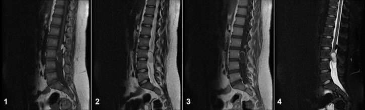
Figure 1-4. Sagittal T1-weighted (1), T2-weighted (2),
postcontrast (3) and STIR (4) images show the tethered cord in level L2-3, the
epidermoid cyst of the filum terminale and the lipoma
DISCUSSION
Improvement in imaging
techniques with widespread use of Magnetic Resonance Imaging (MRI) in
particular, revealed concomitant presence of stretched spinal cord not only
with occult type dysraphism but also with certain abnormalities including
tumor, trauma, arachnoid and lipomyelomeningocele, dermal sinus and
syringomyelia.4
Being the most common form of occult spinal dysraphism,
spinal lipomas occur by encapsulated accumulation of lipid and connective
tissue particles within the spinal cord. Mostly in relation to dural defect,
they extend from spinal cord to subcutaneous tissue.5 Spinal lipomas
not accompanied with spinal dysraphism compose 1% of overall spinal masses with
similar frequency in males and females.6 Frequently, since they are
located in the posterior region of the cervicothoracic or thoracic spinal cord,
the first sign is the difficulty in walking due to posterior compression.7
Afterwards, progressive weakening and spasticity of the extremities arise.
Having long lasting symptoms in general, the average time for patients to admit
a physician is 3-6 years.8 Progressive nature of the complaints
indicates the gradual enlargement of lipomas. In our case, lacking concomitant
spinal dysraphism and being located in the lumbar region, spinal lipoma had
distinct features. Besides, lack of serious complications other than pain is
considered to be related to the particular location of the lipoma in the much lower
segments of the spinal cord.
Identification of hyperintensity on T1 weighted images
while hypointensity on T2 weighted images as well as suppression of signal
intensities at fat suppression sequences facilitate to differentiate lipomas
from other lesions.9
Lumbar puncture, trauma and congenital factors are the
factors documented to be responsible for the development of epidermoid cysts.10
The ratio of intramedullary tumors was reported to be 29.6% in a series of 680
cases with primary spinal tumor by Lunardi et al.11, 0.95% of which
was identified to be epidermoid cyst. Epidermoid cysts are mostly located in
the thoracic region while lumbar location has been documented to be very rare.12
In this respect, lumbar location of the cyst in our
case is worth noting. Moreover, based on medical background of our patient, we
assumed the likelihood of congenital factors to have a role in the etiology of
the disease.
Epidermoid cysts are associated with non-specific MRI
findings such as appearing isodense or hypodense on T1 weighted images while
showing hyperintensity on T2 weighted images. MRI is valuable in identification
of the borders of the lesion as well as its relation to surrounding tissues.13
Strecthing and restricted mobility of the cord via
disturbing cellular metabolism and neuronal function leads to certain
pathophysiological events responsible for progressive ischemia in the cord.14
Frequently, it is diagnosed in childhood and symptomatic onset in adulthood is
very rare.15 Progressive leg and low back pain, weakness and sensory
loss in the lower extremities, pain in the anorectal region, difficulty in
walking, bladder and intestinal dysfunction, extremity abnormalities and
cutaneous defects compose the clinical picture of the disease.1 In
cases with asymptomatic clinical course until adulthood, heavy lifting,
excessive exercise, lithotomy position, childbearing, long-term sitting and
spinal trauma cause the initiation of the symptoms.16
The clinical picture of our case is compatible with
the adulthood presentation of the disease considering pain and numbness as the
leading complaints and evidence of hypoesthesia in the right L5-S1.
Furthermore, fortunately there was no deformity or incontinence development
related to the disease. There was no history of activity capable of yielding
trauma and stretch in the spinal cord. We consider gradual enlargement of
epidermoid cyst as well as age-dependent alterations in the lumbar colon to be
responsible for the increase in spinal cord stretch.
Notably, our case is important in terms of being the
first example of concomitant presence of epidermod cyst and lipoma in the
literature. We think that in concordance with progress that made in imaging
technique, the number of tethered cord syndrome cases that seen with different
pathology will increase.
In conclusion, having likelihood of developing
secondary to different pathologies, tethered cord syndrome must be considered
in the differential diagnosis of patients who admitted with low back pain and
pain and paresthesia in the legs. Detailed medical history and physical
examination are crucial in such patients since the delay in the diagnosis as
well as erroneous administration of electrotherapy with the misdiagnosis of
mechanical low back pain seem quite possible otherwise.
REFERENCES
1. Haro H, Komori H, Okawa A,
Kawabata S, Shinomiya K. Long-term outcomes of surgical treatment for tethered
cord syndrome. J Spinal Disord Tech 2004;17(8):16-20.
2. Guidetti B, Gagliardi FM.
Epidermoid and dermoid cysts clinical evaluation and late surgical results. J
Neurosurg 1997;47(3):12-8.
3. Koyanagi I, Hida K, Iwasaki
Y, et al. Radiological findings and clinical course of conus lipoma:
Implications for surgical treatment. Neurosurgery 2008;63(2):546-51.
4. Duz B, Gocmen S, Secer HI,
Basal S, Gonul E. Tethered cord syndrome in adulthood. J Spinal Cord Med
2008;31(3):272-8.
5. Naidich TP, Blaser SI,
Delman BN, et al. Congenital Anomalies of the Spine and Spinal Cord: Embryology
and Malformations. In: Atlas SW, editör. Magnetic Resonance Imaging of the
Brain and Spine. Philadelphia, PA: Lippincott Williams and Wilkins,
2002:1527-37.
6. Wilson JT, Shapiro RH, Wald
SL. Multiple intradural spinal lipomata with intracranial extension. Pediatr
Neurosurg 1996;24(3):5-7.
7. Fujiwara F, Tamaki N,
Nagashima T, Nakamura M. Intradural spinal lipomas not associated with spinal
dystraphism: A report of four cases. Neurosurgery 1995;37(5):1212-5.
8. Giuffre R. Intradural
spinal lipomas: Review of the literature (99 cases) and report of additional
case. Acta Neurochir (Wien) 1966;14(2):69-95.
9. Tein RD. Fat suppression MR
imaging in neuroradiology: Tecniques and clinical application. Am J Roentgenol
1992;158(4):369-79.
10. Roux A, Merder E,
Larbrisseau A, Oube LJ. Intramedullary epidermoid cysts of the spinal cord. J
Neurosurg 1992;76(4):528-33.
11. Lunardi P, Missori P,
Gagliardi FM. Fortuna A: Long-term results of the surgical treatment of spinal
dermoid and epidermoid tumors. Neurosurgery 1989;25(1):860-4.
12. Lai SW, Chan WP, Hen CY.
MRI of epidermoid cyst of the conus medullaris. Spinal Cord 2005;43(3):320-3.
13. Visdani A, Savoiardo M,
Balestrini MR, Solero L. Iatrogenic intraspinal epidermoid tumor: Myelo-CT and
MRI diagnosis. Neuroradiology 1989;31(2):273-5.
14. Hays RM, Massagli TL.
Rehabilitation concepts in myelomeningocele. In: Braddom RL, editor. Physical
Medicine and rehabilitation. 2th ed. Philadelphia,W. B. Saunders Company,
2000:1217-8.
15. Gupta SK, Khosla VK,
Sharma BS, Mathuriya SN, Pathak A, Tewari MK. Tethered cord syndrome in adults.
Surg Neurol 1999;52(4):362-9.
16. Pang D, Wilberger JE . Tethered cord syndrome in
adults. J Neurosurg 1982;57(6):32-47.