CASE REPORT / OLGU SUNUMU
Doi: 10.5798/diclemedj.0921.2011.04.0053
Leiomyoma of the breast: A Case report
Memede leiomiyom:
Olgu sunumu
Tülin Yalta1,
Ebuzer Bekar2, Ferruh Balaban3
1 Trakya Univeristy, Faculty of
Medicine, Department of Pathology, Edirne, Turkey
2 Sivas State
Hospital, Department of Pathology, Sivas Turkey
3 Sivas Anadolu Hospital, Department of
General Surgery, Sivas Turkey
Yazışma Adresi / Correspondence:
Dr. Tülin Yalta, Trakya University,
Faculty of Medicine, Department of Pathology, Edirne Turkey
Email: tdeyalta@gmail.com
Geliş Tarihi / Received: 07.10.2011, Kabul Tarihi / Accepted: 12.01.2012
ABSTRACT
Leiomyomas are benign smooth muscle neoplasms that are common in the genitourinary
and gastrointestinal tracts. They can occur anywhere in the body but are rare
in the breast. Here we report a case of leiomyoma in the breast in a 43 years
old woman with histological, immünohistochemical
characteristics and review the literature.
Key words: Leiomyoma, breast, diagnosis
ÖZET
Leiomiyomlar düz kas selim tümörlerinden olup
sıklıkla genitoüriner system
ve gastrointestinal sistemde bulunur. Vücutta
herhangi başka bir yerde de bulunabilir ancak memede görülmesi nadirdir. Bu
yazıda 43 yaşındaki kadında memede oluşan leiomiyom
vakası histolojik ve immunokimyasal özellikleri ile
birlikte literatür eşliğinde sunuldu.
Anahtar
kelimeler: Leiomiyom, meme, tanı
INTRODUCTION
Leiomyoma
most commonly occurs in the uterus, small bowel, esophagus and is a rare benign
non epithelial tumor of the breast.1 They have been reported in both
men and women.2 Its histopathology shows interlacing fascicles of
spindle cells that have no atypia or mitoses.3
CASE REPORT
A 43 years
old woman complaining from a right breast lump was seen at the general surgery
clinic of Sivas Anadolu Hospital. She had no family
history of breast cancer and there were no skin changes or axillary lympadenopathy. Physical examination revealed a
well-defined with
There was no intramammarian
lymph node or axillary lympadenopathy. And then the
lesion was surgically excised.
Macroscopic examination of the lesion showed
well-circumscribed, homogeneous, firm, whitish 5x5x3 cm mass. In histopathological (microscopic) examination the lesion was
composed of fascicles of spindle cells that have no atypia.
Necrosis and mitotic activity wasn’t seen. These spindle cells had ovoid
nuclei, delicate chromatin and small inconspicuous nucleoli with eosinophilic cytoplasm. These cells were immunohistochemically diffuse strong positive with
smooth-muscle actin (SMA) and were stained as red histochemically
with Masson-Trichrome stain. On the basis of these
findings we diagnosed this case as leiomyoma of the breast (Figure 1, 2).
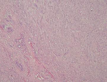
Figure 1. Interlacing bundles of spindle shaped smooth muscle
cells with eosinophilic cytoplasm of the leimyoma nodule at the right side of the figure (HE X50)
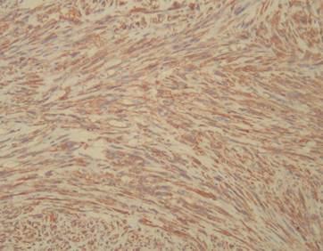
Figure 2. Immunohistochemically
diffuse SMA( smooth muscle actin) positivity of the
spindle shaped cells in the leiomyoma of the breast ( IHC, SMA X100)
DISCUSSION
Leiomyomas are
extremely rare breast tumors.4 First description of this tumor was
made in 1913.5 Most of them occur in subareolar
location.4 We know that these benign pure smooth muscle tumors of
the breast generally occur in middle-aged women from the cases previously
described. The duration of the lesions ranges from 1 month to 26 years and they
involve the right breast more frequently as in our case.6 They also
frequently occur near the nipple and this may be related to the abundance of
smooth muscle cells around the nipple and areola.7 To explain the
origin of these tumors various theories have been proposed. Kaufman et al.
suggested that these neoplasms arise from smooth-muscle cells that surround
capillaries in the subcutaneous tissues of the breast.8 İn the other
hand Diaz-Arias et al. suggested that the origin of these tumors may include
the following;(a) a teratoid origin with extreme
overgrowth of the myomatous elements,(b) embryologically displaced
smooth muscle from the nipple,(c) angiomatous smooth
muscle,(d) multipotent mesenchymal
cell, (e) myoepithelial cells.5
The histopathologic features
of breast leiomyoma are the same as the leiomyomas of
the other sites. Microscopic examination shows groups of interlacing bundles of
spindle shaped cells with eosinophilic cytoplasm.8
Most of them are immunohistochemically positive for vimentin, desmin and
α-smooth muscle actin.5
Fibroadenoma, myoepithelioma, phyllodes tumor and leiomyosarcoma
are in the differential diagnosis of the breast leiomyoma.3 Leiomyosarcoma is the most important differential diagnosis
of this tumor. Histopathologically leiomyosarcomas show cytologic atypia, 2-16 mitotic figures per 10 high-power fields with
atypical mitoses, vascular invasion and necrosis.9 Recommended
treatment of these lesions is complete excision.7
One study indicated that tamoxifen
promotes formation of parenchymal leiomyomas of the
breast.7 Also antiobesity
drugs such as sibutramine and orlistat
may promote the formation of these tumors.4
In conclusion, leiomyoma of the breast parenchyma is a
rare benign neoplasm that is similar to especially fibroadenoma
on ultrasonographic examination. Histopathological
examination and immunohistochemical stains help to
distinguish leiomyoma from other benign and malignant breast lesions.
REFERENCES
1. Koirala K, Shrestha ML, Chalise PR, Shrestha BB, Shrestha R. Leiomyoma of breast: a report of rare case.
Nepal Med Coll J 2008;10(3):
207-8.
2. Ende L, Mercado C, Axelrod D, Darvishian
F, Pascale L, Cangiarella J. Intraparenchymal
leiomyoma of the breast: a case report and review of the literature. Ann Clin Lab Sci 2007;37(3): 268-73.
3. Kotsuma Y, Wakasa K, Yayoi E, Kishibuchi M, Watatani M,
Sakamoto G. A case of leiomyoma of the breast. Breast
Cancer 2001; 8(2): 166-9.
4. Pourbagher A, Pourbagher MA, Bal N, Oguzkurt L, Ezer A. Leiomyoma of the breast parenchyma. AJR 2005;
185(11):1595-7.
5.
Diaz-Arias AA, Hurt MA, Loy TS, Seeger RM, Bickel JT. Leiomyoma of the breast.
Hum Pathol 1989; 20(3):396-9.
6. Sidoni A, Luthy M, Bellezza G, Consiglio MA, Bucciarelli E. Leiomoma of the
breast: case report and review of the literature. The Breast 1999; 8(2):289-90.
7. Son EJ,
Oh KK, Kim EK, Son HJ, Jung WH, Lee HD. Leiomyoma of the breast in a
50-year-old woman receiving tamoxifen. AJR 1998;
171(11): 1684-6.
8. Kaufman
HL, Hirsch EF. Leiomyoma of the breast. J Surg Oncol 1996; 62(1):62-4.
9. Nielsen
BB. Leiomyosarcoma of the breast with late dissemination. Virchows
Arch A Pathol Anat Histopathol 1984;
403(2):241-5.